
First generation cartilage cell implantation (autologous chondrocyte implantation, ACI, autologous chondrocyte transplantation ACT)
Autologous cartilage cell implantation (ACI) is a novel method of treatment for focal cartilage defects. Cells are removed and isolated from non weight bearing articular surface cartilage, and following a longer laboratory culturing cells a reimplanted to the site of cartilage damage producing new cartilage tissue.
Using this relatively novel technique medium and large size chondral defect may be repaired (2-10cm2). The underlying principle of the method is that on site of the cartilage defects cartilage cells embedded in the extracellular matrix have only limited repair capacity. If cells are removed, extracellular matrix is digested, cells are isolated and cultured under regular tissue culture conditions cells demonstrate a good reproductive capacity and cell number will grow rapidly. Once the aimed cell number is reached cell suspension is injected under a flap attached on the top of the cartilage defect and sealed in a waterproof manner. Cells would adhere and start producing the extracellular matrix.
Removal of the small cartilage specimens is done arthroscopically when a few rice body sized cartilage is collected in a sterile tube containing special transfer medium. Specimen is sent to the lab where immediate cell isolation and culturing is commenced. After 4-8 weeks cells would reach the appropriate number for cartilage repair.
A second surgery is performed with mini arthrotomy, the base of the cartilage defect is prepared, periosteal or artificial collagen flap is sutured and sealed with fibrin glue to the edge of the defect. Leaving the uppermost corner of the defect open the surgeon injects the cell suspension under the flap and seals the open corner.
The biggest advantage of the technique is that larger defect sizes may be repaired, and no severe donor site morbidity would occur.
Postoperatively strict rehabilitation must be conducted by experienced physiotherapist, 6 weeks of partial weight bearing with crutches is followed by regular physio sessions. Full recovery should not be expected before 6 months postoperatively.
Drawbacks of the technique is that two surgical intervention is necessary, the freshly developed cartilage is only hyaline-like tissue histologically and is fairly expensive. The laboratory culturing is in the range of 2-4000€.
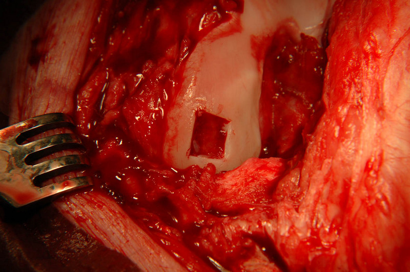 Cartilage defect created on the femur
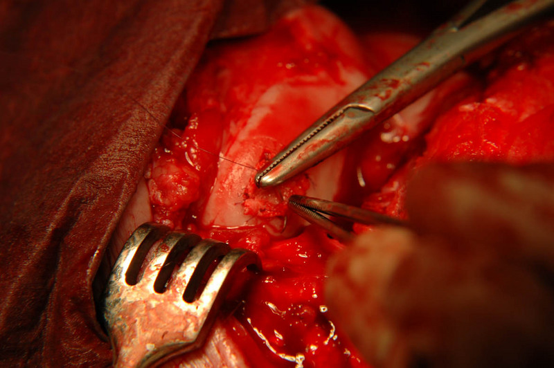 attachment of the periosteal flap on the site of the defect
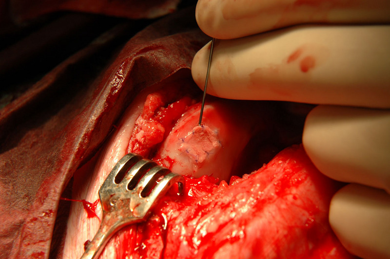 Injection of cartilage cells under the periosteal flap